


How Ultrasound Provides Safe and Accurate Medical Imaging
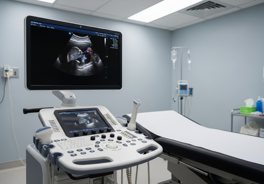
Ultrasound is a diagnostic imaging technique that uses high-frequency sound waves to create real-time pictures of the inside of the body. It is safe, non-invasive, and widely used in healthcare across Australia to examine organs, tissues, and blood flow without radiation exposure.
Unlike X-rays or CT scans, ultrasound imaging does not use ionising radiation, making it a trusted choice for pregnant women, children, and patients requiring repeated scans. Its accuracy in capturing soft tissue structures such as the liver, kidneys, and reproductive organs makes it an essential tool in modern medicine.
Features of Ultrasound Imaging
Ultrasound technology includes distinct features that contribute to its popularity in healthcare.
| Feature | Description | Example |
|---|---|---|
| Non-invasive | No incisions or needles are needed | Abdominal scan for liver evaluation |
| Radiation-free | No ionising radiation involved | Pregnancy monitoring |
| Real-time imaging | Captures moving structures | Foetal heartbeat or blood flow |
| Portable machines | Small and mobile devices | Bedside scans in hospitals |
| Doppler capability | Measures blood flow and velocity | Detecting clots in leg veins |
These features allow clinicians to evaluate conditions accurately while ensuring patient comfort and safety.
Functions of Ultrasound
The main function of ultrasound in Brisbane is to create visual images of internal body structures using sound waves. The transducer sends sound waves into the body, which bounce off tissues and organs, returning echoes that the machine converts into images.
Functions extend beyond imaging. Ultrasound guides interventional procedures, such as biopsies or injections, by providing real-time visuals to improve precision. Doppler ultrasound adds another function by assessing blood circulation, which is critical in identifying vascular conditions like deep vein thrombosis.
Use Cases of Ultrasound
Ultrasound scans are applied in multiple areas of healthcare.
-
Pregnancy and Obstetrics – Monitoring foetal growth, heartbeat, and development.
Example: A 12-week ultrasound shows the baby’s size and confirms gestational age. -
Abdominal Imaging – Examining liver, gallbladder, pancreas, and kidneys.
Example: Detecting gallstones in the gallbladder. -
Musculoskeletal – Assessing muscles, tendons, and joints for injury.
Example: Identifying a rotator cuff tear in the shoulder. -
Cardiac (Echocardiography) – Evaluating heart valves and function.
Example: Detecting mitral valve regurgitation. -
Vascular – Checking blood vessels for clots or blockages.
Example: Detecting deep vein thrombosis in the leg. -
Guided Procedures – Supporting safe needle placement in biopsies.
Example: Breast tissue biopsy for suspected abnormalities.
Pros and Cons of Ultrasound
Every medical tool has strengths and limitations.
| Pros | Cons |
|---|---|
| Safe for all ages including pregnancy | Images may lack clarity in obese patients |
| Cost-effective compared to CT or MRI | Limited penetration through bone or gas-filled organs |
| Real-time imaging for dynamic structures | Operator skill influences accuracy |
| Widely available in clinics and hospitals | Not suitable for all diagnostic needs |
Ultrasound remains a first-line choice due to its safety profile and versatility despite certain limitations.
Target Audience for Ultrasound Services
Ultrasound imaging is relevant to different groups within the healthcare system.
-
Pregnant women: Monitoring foetal development.
-
Athletes: Diagnosing tendon or ligament injuries.
-
Patients with abdominal pain: Identifying gallstones, kidney stones, or liver disease.
-
Cardiac patients: Evaluating heart structure and function.
-
Elderly individuals: Checking for vascular blockages or aneurysms.
Each audience benefits from the non-invasive and radiation-free nature of ultrasound.
Situational Relevance of Ultrasound
Ultrasound becomes highly relevant in specific scenarios:
-
Emergency settings: A trauma patient with internal bleeding can be assessed quickly with an abdominal ultrasound.
-
Pregnancy care: Regular scans provide reassurance and medical insight during prenatal visits.
-
Sports medicine: Rapid identification of tendon or ligament injuries keeps athletes on track with recovery.
-
Vascular concerns: A patient with leg swelling can undergo a Doppler scan to rule out clots.
These examples highlight how ultrasound adapts to different healthcare environments, providing quick, safe, and accurate answers.
Why Ultrasound Is Safe
Ultrasound is safe because it uses sound waves rather than ionising radiation. The absence of radiation exposure makes it suitable for repeated use, even in sensitive cases such as pregnancy or paediatric care.
Scientific studies confirm ultrasound as a safe diagnostic tool with no long-term side effects documented when used within medical guidelines. Its non-invasive approach also reduces risk compared to procedures requiring surgical access.
Accuracy of Ultrasound Imaging
Ultrasound delivers accurate images of soft tissues, vascular structures, and moving organs. Doppler ultrasound enhances accuracy further by showing blood flow direction and velocity.
Accuracy depends on factors such as operator expertise, patient body type, and machine quality. For soft tissue imaging and guiding procedures, ultrasound accuracy exceeds 90% when performed by trained specialists.
In comparison, CT scans and MRI may offer higher detail in some cases, but ultrasound’s combination of safety, speed, and accuracy makes it indispensable.
Comparison of Ultrasound with Other Imaging Techniques
| Imaging Method | Radiation Exposure | Best For | Limitations |
|---|---|---|---|
| Ultrasound | None | Soft tissues, blood flow, pregnancy | Limited view in bone and air-filled areas |
| X-ray | Yes | Bones, fractures | Not suitable for soft tissue detail |
| CT Scan | High | Cross-sectional images of organs | Significant radiation exposure |
| MRI | None | Detailed soft tissue imaging | Expensive and time-consuming |
This comparison shows how ultrasound complements other imaging techniques by filling specific diagnostic roles safely.
visit: https://www.qldradiologyspecialists.com.au/
Conclusion
Ultrasound provides safe and accurate medical imaging by using sound waves to capture real-time views of internal structures without radiation. It offers unique features such as portability, Doppler capability, and real-time visualisation, making it suitable for pregnancy monitoring, musculoskeletal injuries, cardiac assessments, and guided procedures.
Its pros, including safety, affordability, and wide availability, outweigh its limitations, especially when accuracy exceeds 90% in many diagnostic areas. Ultrasound serves diverse audiences, from pregnant women to athletes, and proves situationally relevant across emergency care, sports medicine, and chronic disease management.
Compared to other imaging methods, ultrasound stands out as a trusted diagnostic tool in Australian healthcare. It empowers patients and clinicians with safe, timely, and accurate information, ensuring effective decisions for treatment and care.
Categorii
Citeste mai mult
We love playing Coin Master. The thrill of spinning and winning keeps us hooked. At Khelraja, we enjoy a variety of games, from casinos to slots and betting. But Coin Master holds a special place for us. Coin Master Free Spins are our secret weapon for faster progress and bigger rewards. They give us extra spins without spending money. This means more chances to build villages, attack rivals,...

According to a recent comprehensive by MarkNtel Advisors Turkey Activated Carbon Market research report, the Turkey Activated Carbon market is set for significant growth, driven by factors such as market size, share, and evolving trends. This detailed report offers crucial insights into the market, covering key aspects such as market segmentation and definitions. It outlines the...

"Executive Summary Fertility Services Market : During the forecast period of 2025 to 2032 the market is likely to grow at a CAGR of 7.70%, primarily driven by the anticipated launch of advanced fertility therapies and innovative treatment options DBMR team is focused on understanding client’s businesses and its needs so that the finest market research report is sent to...
The Saudia Airlines ticket booking office in Houston serves travelers flying between Houston and Saudi Arabia with comprehensive support for reservations, ticketing, and travel assistance. Acting as the regional hub for Saudia operations in Texas, it provides essential services to passengers and ensures efficient handling of flight-related queries. Location & Contact Information...
تصوير فعاليات احترافية: سر نجاح المؤتمرات والمعارض والمناسبات المؤسسية في عالم مليء بالمنافسة والانطباعات السريعة، لم يعد نجاح الحدث يُقاس فقط بعدد الحضور أو جودة التنظيم، بل أصبح التوثيق الاحترافي جزءًا أساسيًا من أي مؤتمر، معرض، أو فعالية مؤسسية.إذا كنت تخطط لفعالية في الرياض، جدة، الدمام، أو أي مدينة سعودية، فإن الاستثمار في تصوير فعاليات احترافي لم يعد خيارًا، بل...